On March 9th, Nature published online a research paper titled “Protomelission is an early dasyclad alga and not a Cambrian bryozoan”, co-authored by Associate Professor Lan Tian (co-first author) of the Center for Paleobiology Research of the College of Resource and Environmental Engineering of GZU and Professor Zhang Xiguang and Researcher Yang Jie from Yunnan University. This is the first paper published in the journal Nature in which GZU is listed as a main participating institution.
The study revealed that fossils previously thought to be Cambrian moss animals were actually algae of the early Dasycladales. It has been a long-standing question for evolutionary biologists whether the major phyla of the modern biosphere had already appeared during the Cambrian burst of evolution. Many biological phyla lack hard structures that can be preserved as fossils, but organisms with exoskeletons, such as moss animals, have high fossil preservation potential. Finding moss animal fossils from the Cambrian was a dream for paleontologists seeking evidence to verify the early differentiation hypothesis of the major phyla of organisms. In 2021, a domestic research team published a paper in Nature based on the discovery of a “potential moss animal” from a layer of strata slightly below the Chengjiang fossil group in southern Shaanxi, combined with five similar fossils found in Australia. It is worth noting that the front wall of these specimens’ “worm chambers” is mostly missing, and the identification of the irregular opening as the orifice through which moss animals extend their tentacles is subject to questioning. The orifice is one of the key hard structures of moss animals, and the lack of this structure could shake the conclusion that moss animals existed in the Cambrian.
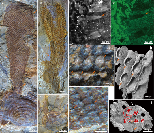
The new Nature paper is based on the analysis of non-mineralized anatomy in Protomelission-like macrofossils from the Xiaoshiba Lagerstätte. The study found that their microscopic structure is very similar to that of previously reported Cambrian phosphatic microfossils resembling moss animals (Figure 1), so the Xiaoshiba specimens were identified as the same lower-level taxonomic unit as the previous ones. On the Xiaoshiba specimens, the leaf-like flanges of the “worm chambers” were found, which suggest a structure different from the tentacle structure of moss animals and belong to the characteristics of the Dasycladales algae. The algal body of the Xiaoshiba algae is covered by a thin and tough surface layer, and once the layer is broken, the residual structure is similar to the “orifice” of the moss animal mentioned earlier (Figure 1f,h); the internal view of the algal body shows that all the algal modules are arranged regularly with sub-diamonds and tiny pores (Figure 1g,i). These comparable characteristic features indicate that the “potential moss animals” from southern Shaanxi and Australia should be early Dasycladales green algae. Therefore, further evidence of moss animal fossils in the Cambrian is still waiting to be discovered.

a-c. Fossils of class Protomelission, originated from the Xiaoshiba Lagerstätte, are often fixed to the shells of other animals with holdfasts (white arrow). The columnar algal body is buried and flattened, showing soft spine-like protrusions in a strip pattern (orange arrow). d, e. Fluorescence and EDX imaging of the details of the protrusions (orange arrow). f-i. Comparison of the details between Xiaoshi green algae and the phosphatized “moss animal”: f, h, the regularly arranged algal body modules, covered with a tough membrane (orange arrow), which becomes an irregularly shaped open hole after it is damaged (indicated by a star), which is not the key structural opening of the “moss animal chamber” (Zhang et al., 2021). g, i, when the body wall of the columnar algal body (or “moss animal” body) on one side is peeled off (white arrow), the rhombic modules on the inner surface of the other side of the algal body are arranged regularly, along with small holes between them (orange arrow).
Editor: Han Xiaomei
Chief Editor: Zhang Yajun
Senior Editor: Yang Nan
Translator: Jia Haibo